
You wake up with an itchy patch on your arm. Is it eczema? A bug bite? Psoriasis?
Without a clear answer, you might use the wrong cream or panic over nothing.
So let’s settle this.
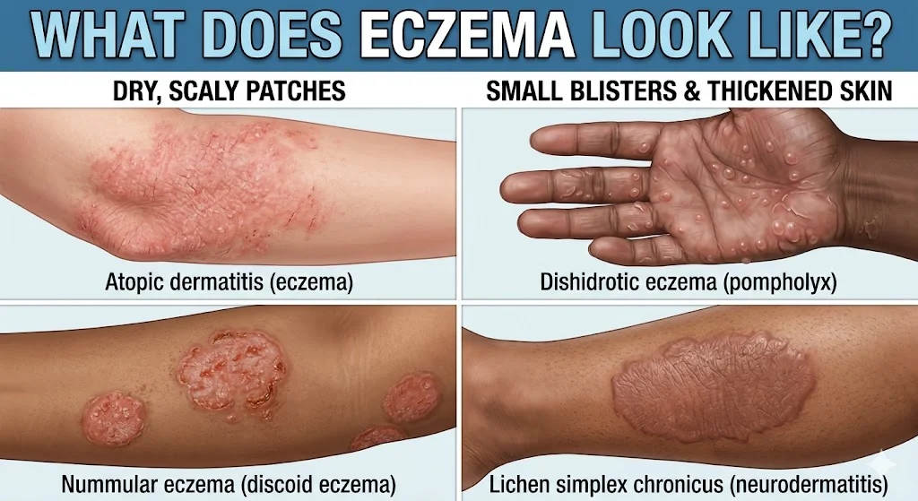
What does eczema look like in real life? Not on airbrushed models. Not in blurry stock photos. But on real people, real skin, and in real flare-ups.
This guide walks you through every visual sign. You’ll learn to spot eczema early, tell it apart from other rashes, and recognize when it’s getting worse. No fluff. Just useful details.
The Five Universal Signs of Eczema: No Matter the Type
Before we break down different kinds of eczema, let’s start with what they all share.
If you see these five features, you’re likely looking at eczema.
Dryness that moisturizers can’t fix
Normal dry skin improves within a day of applying lotion. Eczema doesn’t. The skin feels rough, almost like sandpaper. Cracks form easily. Even thick creams only help for a few hours.
Itching that shows up before the rash
This is a weird one. Many people feel intense itching on a spot that still looks completely normal. The itch arrives first. The redness follows hours or even a day later.
Red or dark patches (color depends on your skin tone)
On fair skin, eczema looks pink or red. On olive skin, it turns purplish. On dark skin (Brown to Black), eczema appears dark brown, gray, or even ashen. The color alone never tells the whole story.
Small bumps that leak when scratched
Look closely. Eczema often starts as tiny, pinhead-sized bumps. They aren’t whiteheads or pimples. These bumps fill with clear fluid. Scratch them, and they weep. That oozing is a major clue.
Thick, leathery skin from chronic scratching
Have you scratched the same spot for weeks? The skin fights back. It grows thicker and darker. The normal skin lines become more visible, almost like tree bark. Doctors call this lichenification.
If you have all five signs, you almost certainly have eczema. If you only have two or three, keep reading. Other conditions can mimic parts of this picture.
Side-by-Side: Six Types of Eczema and What Each One Actually Looks Like
This is where most guides get vague. They say “eczema looks different” but never show you how.
The table below fixes that. Use it like a visual cheat sheet.
| Type of Eczema | Primary Appearance | Color Notes | Common Location |
|---|---|---|---|
| Atopic dermatitis | Dry, scaly patches; sometimes oozing; skin feels rough | Red on light skin; dark brown or purple on dark skin | Elbows, knees, neck, cheeks (babies), wrists |
| Contact dermatitis | Sharply bordered rash; looks exactly where an irritant touched you | Bright red; sometimes with tiny white blisters | Hands (soap), face (makeup), legs (detergent) |
| Dyshidrotic eczema | Deep, tapioca-like blisters; no redness between blisters initially | Flesh-colored or slightly yellow; later turns red and peels | Palms, fingers, soles of feet |
| Seborrheic dermatitis | Greasy, yellowish-white flakes; underlying skin looks swollen | Pink or red; scalp may look darker than surrounding skin | Scalp, eyebrows, sides of nose, behind ears |
| Nummular eczema | Coin-shaped circular spots; size of a dime to a half-dollar | Red, brown, or pink; edges are darker than the center | Arms, legs, torso (rarely on face) |
| Stasis dermatitis | Skin looks thin, shiny, and stretched; brown discoloration around ankles | Reddish-brown; later turns dark purple or rust-colored | Lower legs, around ankles, tops of feet |
Now let’s go deeper. Each type has unique details you won’t find in a basic table.
Atopic Dermatitis: The Most Common Type
This is what most people mean when they say “eczema.”
It starts in childhood for 60% of people. But adults can get it too.
What it looks like during a mild flare:
- Dry, scaly patches the size of a quarter or smaller
- Skin feels like fine sandpaper
- Slightly raised edges
- No oozing or crusting
What it looks like during a severe flare:
- Large patches covering entire elbows or backs of knees
- Deep cracks (fissures) that bleed when you bend
- Clear or yellow fluid weeping from open spots
- Crusty yellow honey-colored scabs (sign of possible infection)
A fact you won’t hear often: Atopic dermatitis almost never appears in the diaper area. If you see a rash on a baby’s bottom that looks like eczema, it’s probably a different condition like a yeast infection or irritant dermatitis.
Contact Dermatitis: The Rash with a Memory
This type follows a simple rule: wherever the irritant touched, the rash appears.
Common triggers include nickel (jewelry), poison ivy, latex, fragrances, and preservatives.
What it looks like:
- Sharp, straight lines or geometric shapes (think watchband rash)
- Blisters in a cluster, not spread out
- Burning sensation often stronger than itching
- Skin feels hot to the touch
Timeline helps you identify it:
- 6 to 48 hours after contact: redness appears
- 2 to 4 days: blisters peak and may ooze
- 1 to 3 weeks: healing begins if you stop exposure
Contact dermatitis looks almost identical to poison ivy reactions. The difference? Poison ivy spreads oils, so you see linear streaks from scratching. Other contact rashes stay put.
Dyshidrotic Eczema: The Tapioca Blister Rash
This type shocks people because it looks nothing like typical eczema.
No dryness. No scaling. Just blisters.
What it looks like:
- Clusters of deep-seated blisters
- Each blister is 1–2mm across
- No redness between blisters (early stage)
- Blisters look like small grains of rice under the skin
What happens next:
After one to three weeks, the blisters dry out. The skin peels in large sheets. Then the area becomes red, cracked, and painful. Eventually it heals, but the skin stays dry for weeks after.
Trigger fact: 50% of dyshidrotic eczema cases link to stress. The other half links to seasonal allergies, humidity changes, or nickel in food (chocolate, nuts, canned goods).
Seborrheic Dermatitis: The Flaky Scalp Rash
People confuse this with dandruff constantly. But dandruff doesn’t cause redness or inflammation. Seborrheic dermatitis does.
What it looks like on the scalp:
- Greasy, yellowish-white flakes
- Flakes stick to the hair and scalp (dandruff falls off easily)
- Red or dark patches underneath the flakes
- No hair loss unless you scratch aggressively
What it looks like on the face:
- Redness in the “butterfly area” (sides of nose, eyebrows, between eyes)
- Greasy scales along the hairline
- Behind the ears: cracks and yellow crusts
Babies get a version called cradle cap. It looks like thick, yellowish-brown scales on the scalp. It rarely itches. Most cases clear up by 12 months.
Nummular Eczema: The Coin Rash
This one earns its name. “Nummular” means coin-shaped in Latin. And that’s exactly what you’ll see.
What it looks like:
- Perfect circles or ovals
- Sharply defined edges (unlike atopic dermatitis which blends into normal skin)
- Multiple spots that don’t merge together
- Spots range from 1cm to 5cm across
The itch cycle is brutal: Nummular eczema itches intensely at night. You wake up scratching. Then the spots weep and crust. Then they itch again. This cycle can last months without treatment.
Key clue: Nummular eczema rarely appears on the face or scalp. If you have coin-shaped spots on your arms or legs but your face is clear, lean toward nummular eczema.
Stasis Dermatitis: The Leg Rash
This type tells you something about your circulation, not just your skin.
Stasis dermatitis happens when leg veins can’t pump blood back to the heart effectively. Fluid leaks into the skin, causing inflammation.
What it looks like (early stage):
- Swollen ankles that look puffy
- Skin feels tight and shiny
- Reddish-brown discoloration around the ankles
- Itching that feels deep, not surface-level
What it looks like (late stage):
- Skin becomes hard and thick (lipodermatosclerosis)
- Dark purple or rust-colored patches
- Small white scars where healed sores used to be
- Open ulcers (venous stasis ulcers) near the inner ankle
Who gets it: Adults over 50, people with varicose veins, those who stand for long hours daily, and anyone with a history of blood clots.
Location Guide: What Eczema Looks Like on Different Body Parts
Eczema doesn’t look the same everywhere on your body. The same person can have dry patches on their arms and oozing blisters on their hands. Location changes the visual story.
Face and Eyelids
Facial eczema is cruel. You can’t hide it. And the skin here is thin, so inflammation looks worse.
What it looks like on cheeks:
- Diffuse redness or darkening
- Rough, sandpaper texture
- Fine white flakes, not greasy scales
- Warm to the touch during flares
What it looks like on eyelids:
- Swollen, puffy folds
- Red or dark purple discoloration
- Skin looks wrinkled and crepey
- No flaking (eyelids don’t produce flakes like the scalp)
A critical warning: Never use steroid creams on your eyelids without a doctor’s guidance. Eyelid skin absorbs medication six times faster than forearm skin. Overuse leads to glaucoma and thinning skin.
Hands
Hand eczema deserves its own category. Your hands touch everything. Soaps, sanitizers, food, water, gloves. All of it triggers flares.
What it looks like:
- Dry cracks on fingertips (painful and slow to heal)
- Redness that stops sharply at the wrist
- Small blisters along the sides of fingers
- Nail changes (ridges, pitting, thickening)
- The “ring sign” – dryness around a finger like a tight ring left an imprint
Shocking fact: Healthcare workers have a 30% lifetime risk of developing hand eczema. Constant handwashing strips the skin barrier. Within two years of starting nursing or dental hygiene, most see their first flare.
Neck
The neck moves constantly. That motion irritates eczema further. You also scratch your neck in your sleep without realizing it.
What it looks like:
- Thick, leathery patches (lichenification)
- Horizontal lines that look like wrinkles but aren’t age-related
- Darker skin than the surrounding area
- Hair follicles become more visible (follicular accentuation)
Neck eczema often signals an airborne allergy. Pollen, pet dander, dust mites, and mold land on your neck throughout the day. If your neck flares but your arms stay clear, consider environmental triggers.
Legs
Leg eczema splits into two patterns: front of the shins or back of the knees.
Front of shins:
- Dry, cracked, “impulsive pavement” appearance
- Sharply defined oval patches
- Bleeding from minor scratches
- Slow healing (shins have poor blood flow)
Back of knees:
- Moist, red, oozing patches
- Skin feels soft and macerated from sweat
- Dark brown discoloration from chronic inflammation
- Thickened folds
Leg eczema also gets infected more often than other locations. Bacteria and fungi thrive in the warm, moist knee creases. Watch for yellow crusts or spreading redness.
Scalp
Scalp eczema is almost always seborrheic dermatitis. But atopic dermatitis and contact dermatitis can also affect the scalp.
What seborrheic dermatitis looks like on the scalp:
- Greasy yellow-white flakes stuck to the hair shafts
- Red or dark patches between flakes
- No hair loss (this is key – scarring alopecia looks different)
- Flakes return within days of washing
What contact dermatitis looks like on the scalp:
- Itching within hours of using a new hair dye or shampoo
- Red, swollen, oozing patches
- Blisters in severe cases
- Flakes are dry, not greasy
Important distinction: Scalp psoriasis has thick, dry, silver-white scales. Eczema scales are smaller and yellower. Psoriasis plaques feel like candle wax. Eczema feels like dry cereal flakes.
Baby Cheeks and Chin
Infant eczema follows a predictable pattern. It starts on the cheeks and chin around two to four months of age.
What it looks like:
- Red, raw, weeping patches on both cheeks
- Chin looks irritated, especially after drooling or spitting up
- Scalp may have cradle cap (seborrheic dermatitis)
- The diaper area stays clear
Babies can’t control scratching. They rub their face on the mattress or use their hands. This creates a “shiny” appearance on the cheeks from constant friction.
Timeline fact: 50% of babies with eczema outgrow it by age five. Another 30% outgrow it by puberty. Only 20% carry eczema into adulthood.
Critical Comparison: Eczema vs. Look-Alikes
What does eczema look like compared to other rashes? This section saves you from treating the wrong condition.
Eczema vs. Psoriasis
| Feature | Eczema | Psoriasis |
|---|---|---|
| Scale color | Yellow-white or off-white | Silver-white |
| Scale texture | Fine, flaky, like dandruff | Thick, dry, like mica flakes |
| Itch intensity | Extreme, often the first symptom | Mild to moderate |
| Bleeding when scratched | Yes, easily | No, but you may see pinpoint bleeding (Auspitz sign) |
| Common age of onset | Infancy or childhood | 15-35 years or 50-60 years |
| Nail changes | Ridges, pitting, thickening | Pitting, oil spots, crumbling |
Eczema vs. Fungal Infection (Ringworm)
Ringworm is the most common eczema imposter. Here’s how to tell them apart.
Ringworm signs:
- A single spot or just a few spots (eczema has many)
- Clearing center with an active red border
- Raised, scaly edge
- No weeping or oozing unless infected
- Spreads outward in a circle over weeks
Eczema signs:
- Multiple spots that merge together
- Even redness across the whole patch
- Intense itching all the time, not just at night
- History of dry skin or allergies
The scalpel test: Doctors scrape the edge of the rash and look under a microscope. Fungal hyphae (threads) confirm ringworm. No hyphae suggests eczema.
Eczema vs. Allergic Reaction (Hives)
Hives come and go quickly. Eczema stays put.
| Feature | Hives | Eczema |
|---|---|---|
| Duration | Each welt lasts <24 hours | Each patch lasts weeks or months |
| Movement | Welts disappear in one area and appear elsewhere | Stays in the same spots |
| Appearance | Smooth, raised, no scale | Rough, dry, scaly |
| Color | Pale center with red border | Uniform red or dark |
| Response to antihistamines | Dramatic improvement | Minimal improvement |
Eczema vs. Scabies
Scabies is a mite infestation. It mimics eczema so well that doctors misdiagnose it constantly.
Scabies clues:
- Intense itching that gets worse at night
- Tiny burrows (thin gray or silver lines) between fingers, on wrists, or along belt lines
- Multiple family members or close contacts have the same itch
- No response to steroid creams (they actually make scabies worse)
Eczema clues:
- Itching happens any time of day
- No burrows (just bumps or scaling)
- Family members without eczema don’t itch
- Steroid creams help temporarily
One telling sign: Scabies rarely affects the head and neck in adults. If you have an intensely itchy rash on your face that looks like eczema, it’s probably not scabies.
Skin Tone Matters: What Eczema Looks Like on Dark Skin vs. Light Skin
Most eczema photos show light skin. That’s a problem.
On dark skin (Fitzpatrick types IV to VI), what does eczema look like changes dramatically.
Color Differences
On light skin (Types I-III):
- Redness is obvious and bright
- Pink or salmon-colored patches
- Inflammation looks “angry”
On dark skin (Types IV-VI):
- Redness turns into dark brown, purple, or ashen gray
- Patches look almost black in severe flares
- Inflammation looks “dusky” or “muddy”
- Surrounding skin may have a lighter halo (peripheral hypopigmentation)
Post-Eczema Changes
After the rash heals, dark skin doesn’t return to normal quickly. Two things happen.
Hyperpigmentation (dark spots):
The inflamed skin produces extra melanin. These dark spots last for months. Sometimes they last more than a year. They look like flat, brown patches exactly where the eczema used to be.
Hypopigmentation (light spots):
Sometimes inflammation destroys melanocytes temporarily. You get white spots that look like vitiligo. These also fade slowly. Sun exposure makes the contrast worse because normal skin tans but the white spots don’t.
Realistic expectation: Even after successful treatment, the color changes take three to twelve months to fade completely. That doesn’t mean the eczema is still active. It means your skin needs time to repigment.
Textural Differences on Dark Skin
Lichenification (thickened skin from scratching) looks more dramatic on dark skin.
The normal skin lines become dark, deep, and pronounced. You can see every crisscross. The skin feels like thick leather or tree bark.
Papular eczema is also more common in dark skin. Instead of large red patches, you get hundreds of small, firm bumps. They look like goosebumps that never go away. Each bump itches intensely.
Stages of an Eczema Flare: From First Itch to Healing
Eczema follows a predictable sequence. Learn these stages so you know what comes next.
1: The Silent Dryness
Your skin feels tight. Maybe it looks slightly dull or ashy. No redness. No bumps. Just a subtle change in texture.
What’s happening: The skin barrier has microscopic cracks. Water escapes. Irritants enter. But inflammation hasn’t started yet.
What to do: Heavy moisturizer within three minutes of bathing. If you skip this stage, you move to stage two.
2: The First Itch Without a Rash
You feel an itch in a specific spot. You look. Nothing’s there. You scratch anyway. Still no redness.
What’s happening: Inflammatory chemicals (histamine, substance P) have been released. But visible blood vessel dilation hasn’t occurred yet.
What to do: Apply a cool compress. Use an antihistamine if the itch is widespread. Do NOT scratch – stage two itching can last hours before the rash appears.
3: Red or Dark Patches Appear
Now you see it. A raised, warm patch. Red on light skin. Dark brown or purple on dark skin. The edges may be blurry or sharp depending on the type.
What’s happening: Blood vessels dilated. Immune cells migrated into the skin. Fluid starts leaking into the tissue.
What to do: Apply a topical steroid (over-the-counter hydrocortisone for mild cases; prescription strength for moderate to severe). Moisturize five times daily.
4: The Wet, Oozing Phase (Acute Flare)
This stage scares people. The skin weeps clear or yellow fluid. Small blisters may appear. Crusting develops overnight.
What’s happening: The skin barrier has completely failed. Fluid from leaking blood vessels reaches the surface. Bacteria may colonize the wet areas.
What to do: Wet wrap therapy. Apply steroid cream, then cover with a damp cotton sleeve or gauze, then a dry layer. Leave for two to six hours. Change twice daily. See a doctor if oozing doesn’t improve within three days.
5: The Chronic Scratch Cycle
You’ve scratched for weeks. The skin doesn’t look red anymore. Instead it looks thick, dark, and leathery. The normal skin lines stand out like topographical map lines.
What’s happening: Repeated scratching caused fibroblasts (skin cells) to multiply and lay down extra collagen. The skin thickened to protect itself.
What to do: This stage is hard to reverse. You need stronger prescription treatments like calcineurin inhibitors (tacrolimus, pimecrolimus) or a mid-potency steroid. Oral antihistamines at night to stop sleep scratching.
6: Healing and Peeling
The redness fades. The itching stops. But now the skin peels in large flakes or sheets.
What’s happening: The top layers of damaged skin are shedding. New skin grows underneath.
What to do: Do NOT pick the peeling skin. Continue moisturizing. Use a gentle exfoliant (like urea cream) to help flakes shed evenly. Protect the new skin from sun – it burns easily.
7: Residual Color Changes
The eczema is gone. But dark or light spots remain. This isn’t active disease. Just a cosmetic remnant.
What’s happening: Melanin distribution was disrupted. It takes time to normalize.
What to do: Sunscreen daily. Sun exposure makes dark spots darker and light spots more obvious. Fading creams (azelaic acid, kojic acid, vitamin C) can speed things up. Most importantly: wait. Time heals color changes.
When to Stop Guessing and See a Doctor
You’ve read this far. You’ve compared photos in your head. You still aren’t sure what does eczema look like on your specific rash. That’s okay. Some cases need a professional.
See a dermatologist if you experience any of these:
- Eczema covers more than 20% of your body. That’s roughly the size of your entire front torso plus one entire arm. Widespread eczema needs prescription treatment.
- Home treatments make it worse. You tried hydrocortisone and thick moisturizers for two weeks. The rash spread or became more painful. That suggests infection or a wrong diagnosis.
- You see honey-colored crusts. That’s not dried eczema fluid. That’s Staphylococcus bacteria. You need oral or topical antibiotics plus steroids.
- Red streaks travel away from the rash. This is lymphangitis – the infection entered your lymphatic system. Go to urgent care today.
- You have eczema and a fever over 100.4°F (38°C). Eczema herpeticatum (herpes virus superinfection) is a medical emergency. It looks like punched-out craters filled with blood or pus. Don’t wait.
- You can’t sleep through the night. Sleep deprivation from itching affects your immune system, mental health, and work performance. A dermatologist can prescribe sedating antihistamines or better topical treatments.
- You’ve been using steroid creams for more than four weeks without a break. You need a doctor to guide tapering and switch you to a steroid-sparing medication.
FAQs
Can eczema look like tiny pimples?
Yes. Dyshidrotic eczema starts as deep, flesh-colored bumps that resemble small whiteheads but without a visible pore.
Does eczema always ooze?
No. Only acute flares with broken skin ooze. Chronic, thickened eczema stays dry.
What does mild eczema look like on hands?
Just dry, slightly red skin between fingers or on knuckles. No blisters. No cracking. It looks like “winter hands” that never improve with lotion.
Is eczema raised or flat?
Usually raised. Inflamed skin swells. Chronic scratching flattens it over time, but the edges stay raised.
What does healing eczema look like?
Peeling skin, less redness, no new bumps, and itching that feels like a dull ache instead of a sharp need to scratch.
Can eczema look like ringworm?
Rarely. Nummular eczema forms coin-shaped spots but never has a clearing center. Ringworm has a central healing zone with an active border.
What does infected eczema look like?
Yellow honey-colored crusts, spreading redness, warmth, swelling, and pain (eczema normally doesn’t hurt – it itches).
Does eczema leave scars?
Not true scars unless you scratched deeply. But color changes (dark or light spots) last for months and look like scars. These fade eventually.
Conclusion:
You don’t need to memorize every detail. Keep this cheat sheet handy instead.
What does eczema look like? One quick summary:
- Dry, rough patches that moisturizers won’t fix
- Itching that starts before the rash appears
- Red (light skin) or dark brown/gray (dark skin) patches
- Small bumps that leak clear fluid when scratched
- Thick, leathery skin from weeks of scratching
What it does NOT look like:
- A single ring-shaped spot with a clear center (that’s ringworm)
- Silver-white thick scales (that’s psoriasis)
- Welts that disappear within 24 hours (those are hives)
- Tiny gray burrows between fingers (that’s scabies)
Trust your eyes. Compare what you see to the descriptions here. When in doubt, see a dermatologist. Eczema is treatable but only if you know what you’re looking at.
Now go check that itchy patch. You know exactly what to look for.
Read More Related Articles:
- What Does QHS Stand For? A Clear Medical Definition In 2026
- MRN Mean in Medical Terms: Medical Record Number Explained In 2026
- ADL Means in Medical Terms: Assessment & Clinical Use In 2026

Neon Samuel is a digital content creator at TextSprout.com, dedicated to decoding modern words, slang, and expressions. His writing helps readers quickly grasp meanings and understand how terms are used in real conversations across text and social platforms.